x rays of bunions
For patients undergoing surgery to repair a bunion deformity of the foot non-weight-bearing x rays taken immediately after surgery can provide a good estimate of the risk that the bunion will. Be sure not to leave it on longer than 20 minutes at a time.
 |
| Pin On Photos |
Bunion pain keeping you from doing what you love.
. Our Los Angeles podiatrists will likely order X-rays to diagnose the progression of the bunion before bunion surgery. Halluces valgi is a fixed abduction of the first metatarsophalangeal joint of the great toeIt is usually due to metatarsus primus varus which is a medial deviation or adduction of the first metatarsal with an increased first-second metatarsal angle. Posted by 6 days ago. An X-ray of the foot reveals how far the big toe joint has moved away from the other toes and whether any other bones in.
Corns or calluses between the toes or smaller bunionettes. Ziba required an osteotomy bunionectomy hammertoes corrections with our Ossio implant and metatarsal phalangeal joint relocation and plantar plate repair. X-rays can make these estimates based on a few simple measurements of the patients hallux valgus angle HVA or the angle formed by the toe. Get a Medical Exam to Identify Risk Stroke Vascular Disease.
Two angles are assessed. A recent study published in The Journal of Bone and Joint Surgery shows that x-rays taken right after bunion surgery can reliably predict the risk of bunion recurrence over time. Doctors departments Print Diagnosis Your doctor can identify a bunion by examining your foot. A few days before the bunion procedure you may need to stop taking certain medications including blood thinners.
It is the lateral counterpart of the more common bunion of the first metatarsophalangeal joint and when they occur together often with spreading of the other metatarsals patients. A normal first metatarsophalangeal angle is. After the physical exam an X-ray of your foot can help your doctor determine the best way to treat it. Orthopedic surgeons diagnose bunions on the basis of a physical examination and X-rays taken while the person is weightbearing standing.
Take Control of Your Health. Her bunion was so severe she had placed a lot of weight on her 2nd and 3rd toes which had become totally dislocated as shown in x-rays. Two angles are assessed. If a bunion causes significant pain or severely changes the shape of the big toe doctors may recommend X-ray imaging.
It is considered abnormal if greater than 1518. Ad Return To Normal Motion Faster With Less Stiffness Compared With Other Bunion Surgeries. Pain around the ball of the foot. A sub to discuss bunions.
The alignment of your foot bones changes when you stand. A sub to discuss bunions. Bunions are generally categorized as mild moderate or severe. The hallux valgus angle HVA is the angle between the longitudinal axes of the proximal phalanx and the first metatarsal bone of the big toe.
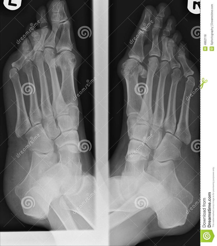
Bunion x ray stock pictures royalty-free photos images. Bunion can be diagnosed and analyzed by projectional radiography X-ray which should be weight-bearing. X-ray of the foot valgus deformity of the toe. 18k members in the bunions community.
A bunionette also known as a tailors bunion or metatarsus quintus varus is a bony prominence at the lateral 5 th metatarsal head. Predicting the Return of Bunions with X-Rays. In some cases arthritis may also be seen. While bunion surgery involves repositioning the malaligned bones most procedures also involve shaving away any bony prominence at the big toe joint.
22k members in the bunions community. They may also draw blood or ask for a urine test to rule out any underlying medical conditions that may cause complications. The big toe pointing in the direction of the smallest toe. Active Bunion Recovery Can Be Faster With Fewer Complications Versus Other Surgeries.
Foot x-ray with an area of pain or swelling - bunion xray stock pictures royalty-free photos images podiatry icons - bunion xray stock illustrations Radiology center France technician carries out a foot x-ray on a patient who had an operation on a hallux valgus. Although your doctor will probably be able to diagnose your bunion based on your symptoms and the appearance of your toe they will also order an X-ray. A bunion is usually not a serious issue but it is a painful condition. More severe bunions restrict movement in the foot.
An angular bony painful bump on the outside base of the big toe sometimes with hardened skin or a callus covering it. More Information X-ray Treatment Treatment options vary depending on the severity of your bunion and how much pain it causes. 10 votes 12 comments. Bunion Diagnosis Bunions are visually apparent.
A hallux valgus plural. X-ray of the feet valgus deformity of the toe or a bone on a finger 3rd degree. An X-ray will allow your doctor to. Swelling redness and pain around the big toe area that can appear shiny.
They may also measure the angles between the bones of the toes. What causes them and what to do about them. Discover the Phantom MIS solution. X-rays can easily identify whether there is a bony enlargement.
Log in or sign up to leave a comment. Not a place for sexualizing feet. To fully evaluate your condition your foot and ankle surgeon may take a foot x-ray to see any abnormal angles between the big toe and the foot to determine the degree of the deformity and assess the changes. Citation DOI article data.
Its common to see bony enlargements that occur with bunions but they also occur when a bunion is absent. The doctor may take x-rays or perform an electrocardiogram to evaluate your heart and lung function. Footwear X-rayed Ladys foot in a high heeled shoe -. To ease swelling and pain wrap a bag of frozen vegetables or crushed ice in a towel and put it on your bunion.
To judge how severe a bunion is clinicians take an x-ray and measure angles between certain bones in the foot in particular the hallux valgus angle HVA the angle between the first metatarsal and the big toe and the angle formed by the first and second metatarsals called the intermetatarsal angle IMA. Radiographic features Plain radiograph. What causes them and what to do about them. A doctor should be able to determine the exact location of a bunion with an x-ray.
X-rays provide images of dense structures such as bone. Check the alignment of your toes and look for damage to the MTP joint. A doctor will measure the angles of the bones of the foot including the big toe. Ad Learn more about new bunion treatments and get back to what you love.
A good foot x. Ziba is a 67-year-old female with years of foot pain. Intermetatarsal angle the angle between the first and second metatarsals the bones that lead up to the bases of the big toe and second toe. Ad Identify Health Risks Before Symptoms Develop.
 |
| Faq S About Bunions Bunion Bunion Surgery Bunion Treatment |
 |
| What Does A Bunion Look Like Bunion Bursitis X Ray |
 |
| Bunions Foot First Podiatry Centers Bunion Podiatry Bunion Surgery |
 |
| Before And After Pictures Of Bunion And Tailor S Bunion Surgery |
 |
| Pin Su Before And After Pics |
Posting Komentar untuk "x rays of bunions"